


1. Introduction
2. Literature Search
3. In Vitro Production
3.1. Ovum pick-up and donor management
3.2. In vitro maturation
3.3. In vitro fertilization
3.4. In vitro culture
3.5. Cryopreservation and transfer
4. Determinants of Efficiency
4.1. Reduction in embryo developmental competence
4.2. Donor factors
4.3. Sire factors
4.4. Recipient factors
5. Quality of In Vitro Produced Embryos
5.1. Phenotypic differences
5.2. Chromosomal integrity
5.3. Epigenetic reprogramming and offspring phenotype
5.4. Challenges associated with embryo transfer
6. Applications in Cattle Breeding
6.1. Accelerating genetic gain
6.2. Sexed semen and beef-on-dairy strategies
6.3. Embryo trade and genetic conservation
7. Emerging Technologies
7.1. Biphasic and physiologically informed maturation
7.2. Embryokines
7.3. Time-lapse imaging
7.4. In vitro breeding
8. Conclusion
1. Introduction
The first calf derived from a transferred embryo was born in December 1950, but for most of the following half-century embryo technology in cattle was limited by two constraints. Superovulation followed by uterine flushing multiple ovulation and embryo transfer (MOET) yields a modest and historically static number of embryos per procedure, currently averaging 8.8 ova and 5.7 transferable embryos per flush, essentially unchanged from two decades earlier [1]. Separately, until dense single-nucleotide polymorphism genotyping became available for cattle in 2009 [2], females could not be ranked with accuracy approaching that achievable for bulls, so multiplying the offspring of a given cow offered limited genetic return.
Both constraints have been relaxed. Genomic evaluation permitted accurate selection of young animals and a large reduction in generation interval [3]. IVP made it possible to convert a genetically superior female into many embryos independently of her reproductive cycle [4]. Their conjunction transformed the industry the number of IVP cattle embryos exceeded the number of in vivo derived embryos for the first time in 2016, and by 2024 more than two million transferable IVP embryos were recorded worldwide against 296,520 in vivo derived, with IVP representing approximately 87% of bovine embryo activity and growing at about 14.2% per year over the past decade [1].
The expansion has been driven less by improvements in laboratory efficiency than by structural advantages. Oocytes can be recovered repeatedly from cyclic females regardless of cycle stage, from cows in early pregnancy, from prepubertal heifers, and from animals with uterine pathology that would preclude flushing. A single ejaculate can be distributed across many fertilization drops, which changes the economics of sexed semen. Fixed laboratory costs are diluted by throughput in a way that flushing cannot match [1,4].
This review describes the technology stage by stage, evaluates the donor, sire, laboratory and recipient factors that determine its outcome, considers the quality of the resulting embryo, and surveys applications in breeding and emerging technical developments. Throughout, direct experimental evidence is distinguished from mechanistic inference, since the two are frequently conflated in this literature.
2. Literature Search
A focused narrative search was conducted in PubMed, web of science and scopus. Search combinations included bovine or cattle with in vitro embryo production, OPU, oocyte maturation, IVF, blastocyst, embryokine, cryopreservation, antral follicle count, embryo transfer (ET) and genomic selection. Priority was given to publications from 2015 onward, with earlier work retained where it is foundational or where the relevant experiment has not been repeated. Industry statistics were taken from the International Embryo Technology Society (IETS) Data Retrieval Committee annual reports. Reviews and meta-analyses were preferred over single primary studies where both addressed the same question, and reference lists of retrieved reviews were screened for additional sources.
Evidence was classified into three categories. Direct evidence required an experiment in which a defined exposure was manipulated and a developmental outcome measured in the same study. Indirect evidence described a molecular or morphological difference without linking it to a developmental endpoint. Descriptive evidence comprised industry statistics and observational reports. The distinction is applied because much of the literature on culture supplementation is indirect while the conclusions commonly drawn from it are causal.
3. In Vitro Production
Bovine in vitro embryo production comprises a sequence of ovum pick-up, in vitro maturation, in vitro fertilization, in vitro culture, cryopreservation, and embryo transfer, with each stage characterized by distinct conditions, efficiencies, determinants, and limitations (Table 1).
Table 1.
Stages of in vitro embryo production, with typical conditions, efficiency, determinants and limitations
3.1. Ovum pick-up and donor management
Transvaginal ultrasound guided follicular aspiration is performed with a probe housed in a rigid guide, through which a needle of 17–19 gauge punctures visible antral follicles under vacuum. Sessions are commonly performed once or twice weekly and can be repeated over long periods without measurable damage to the reproductive tract [4]. Aggregated 2024 data give a useful benchmark: 393,315 donor sessions yielded 7,703,541 oocytes and 2,011,185 transferable embryos, corresponding to approximately 19.6 oocytes and 5.1 transferable embryos per session [1].
Whether to precede aspiration with exogenous follicle-stimulating hormone remains contested, and practice differs sharply by region: 53.9% of reported cycles worldwide were stimulated in 2024, but 94.9% in Europe against 18.9% in Oceania [1]. Stimulation generally increases both the number and the mean competence of recovered oocytes, particularly when combined with a coasting interval, at the cost of gonadotropin, additional handling and a longer inter-session interval. A further shift is that abattoir-derived oocytes now account for under 1% of global IVP output [1] commercial production runs almost entirely on identified, genetically evaluated donors, which reverses the pattern of the 1980s and 1990s and has implications for the generalizability of experiments conducted in slaughterhouse material.
3.2. In vitro maturation
Cumulus oocyte complex (COC) are graded morphologically and cultured for approximately 22–24 h at 38.5 °C in 5% CO₂, most often in TCM-199 supplemented with gonadotropins, serum or a serum substitute, and frequently steroids and growth factors [4]. Maturation comprises two processes that are synchronized in vivo. Nuclear maturation is the resumption of meiosis and arrest at metaphase II. Cytoplasmic maturation is the less visible reorganization of organelles, accumulation of maternal transcripts and protein, redistribution of mitochondria and cortical granules, and establishment of the redox and calcium signalling capacity on which the embryo depends before genome activation [5].
Removing the complex from the follicle abolishes the inhibitory signalling principally cyclic GMP transfer from cumulus cells and the resulting maintenance of oocyte cyclic AMP that sustains meiotic arrest [5,6]. Meiosis resumes prematurely while cytoplasmic maturation lags. Nuclear maturation therefore proceeds efficiently, with 80–90% reaching metaphase II [7], while the oocyte carries a reduced developmental endowment. This uncoupling is the principal mechanistic explanation for the inefficiency of the system as a whole.
3.3. In vitro fertilization
Frozen-thawed semen is separated from extender and damaged cells by discontinuous density gradient centrifugation or swim-up, capacitation is conventionally induced with heparin [8], and gametes are co-incubated for 18–22 h at approximately one million sperm per millilitre. Two features distinguish this from fertilization in vivo. First, sperm numbers reaching the oviduct after insemination are estimated in the tens of thousands and those reaching the site of fertilization probably in the hundreds or fewer [9]; transit imposes non-random selection, and sperm with fragmented DNA are less likely to pass [10]. Second, heparin is an incomplete surrogate for capacitation in the oviduct, which also prolongs sperm survival and contributes to polyspermy control. Co-culture of gametes with oviductal epithelial cells in a microfluidic device reduced polyspermy and parthenogenetic activation relative to conventional IVF, which demonstrates that these conditions are modifiable [11].
3.4. In vitro culture
Presumptive zygotes are denuded and cultured for approximately seven days to the blastocyst stage, most often in synthetic oviductal fluid or a derivative. Two practices are near-universal. Culture at reduced oxygen tension, approximately 5% rather than atmospheric, limits oxidative damage. Group culture outperforms individual culture [12], an effect attributed to embryo-derived paracrine factors and now reconciled with individual tracking through well-of-the-well and microwell dishes [13]. Serum presents a persistent dilemma: it supports development but promotes intracellular lipid accumulation, a principal cause of poor cryotolerance, and alters both the epigenome [14] and the activity of supplemented growth factors [15].
3.5. Cryopreservation and transfer
Embryos are graded on IETS criteria on day 7 and transferred fresh, slow-frozen or vitrified. Cryotolerance has been the practical weak point of the technology, with post-warming survival and pregnancy consistently lower than for in vivo-derived embryos [16]. Three lines of countermeasure exist: reducing lipid accumulation during culture; mechanical delipation, effective but impractical at scale; and improvement of the protocol itself. Freezing in chemically defined medium using polyvinyl alcohol in place of albumin gave pregnancy rates comparable to fresh transfer (50% versus 52%) [17], and an in straw warming protocol permitted direct transfer of vitrified embryos (40% versus 43%) [18]. These advances are visible in practice: frozen or vitrified IVP embryos rose from 10.7% of all bovine transfers in 2015 to 35.1% in 2024 [1].
4. Determinants of Efficiency
4.1. Reduction in embryo developmental competence
Attrition becomes visible during cleavage stage development (Fig. 1), which has encouraged sustained attention to post-fertilization culture. The direct evidence does not support that inference. Rizos et al obtained 39% blastocysts when maturation, fertilization and culture all occurred in vitro, 78% when maturation alone occurred in vivo, and in a second experiment 39%, 58% and 74% for fully in vitro, in vivo maturation only, and in vivo maturation with in vivo fertilization [19]. Gad et al reported 12.2% blastocysts under fully in vitro conditions; transferring in vitro-produced embryos to the uterus at the 16 cell stage did not improve this (10.6%) and transfer to the oviduct at the 4 cell stage improved it only to 26.6%, whereas embryos produced in vivo and then cultured in vitro from the 4 cell or 16-cell stage yielded 83.0% and 69.8% [20].
An embryo generated in vivo therefore tolerates in vitro culture, while an oocyte matured and fertilized in vitro develops poorly wherever it is subsequently placed. The same conclusion has been reached in other mammalian species [21]. Culture conditions are not without effect they demonstrably shape blastocyst quality, gene expression and epigenetic state but they are not the quantitative bottleneck, and interventions applied after fertilization are constrained by a ceiling set earlier.
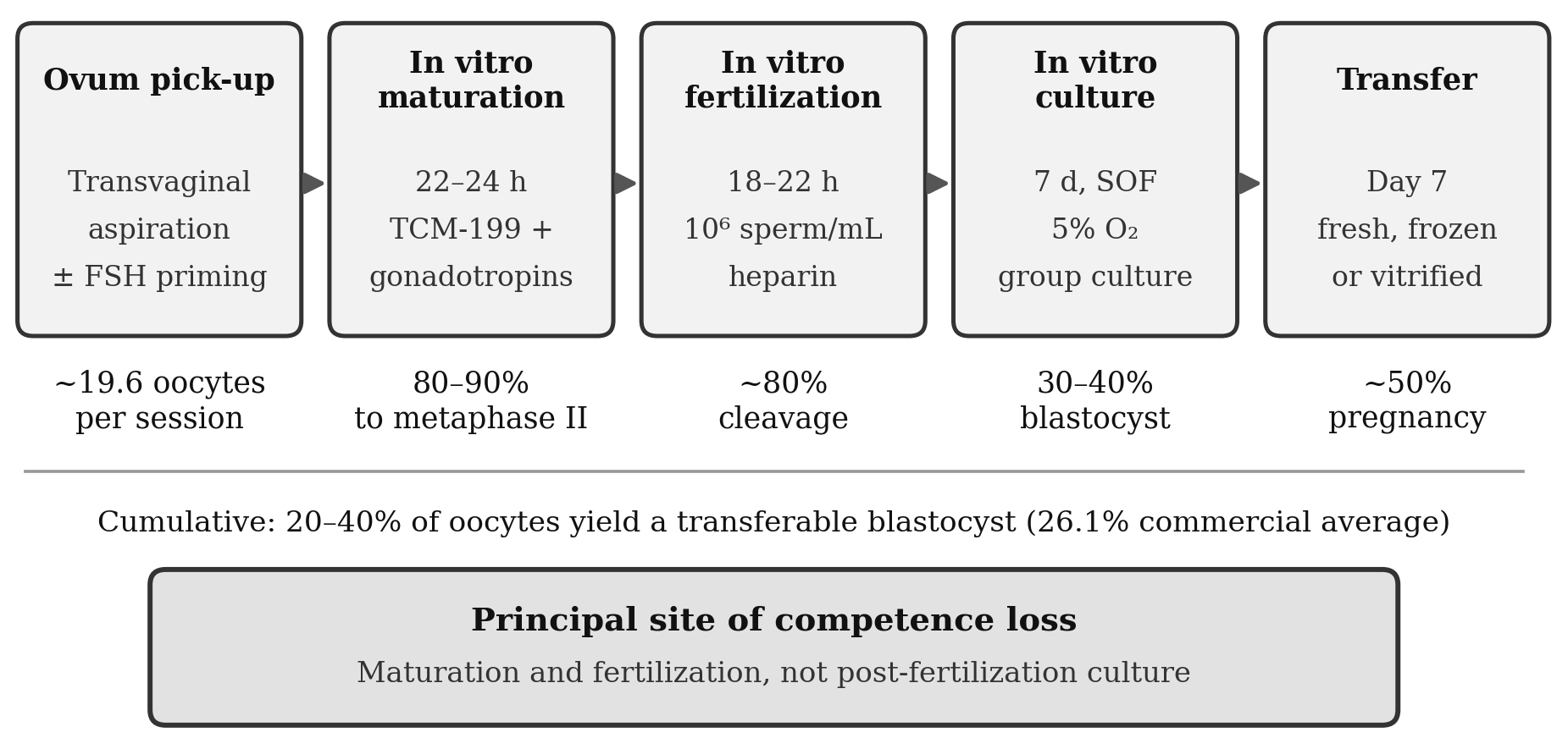
Fig. 1
Bovine in vitro embryo production sequence with typical stage wise efficiency. Nuclear maturation and cleavage proceed at high nominal rates, and attrition becomes visible during cleavage stage development, but environment-partitioning experiments locate the loss of developmental competence in maturation and fertilization rather than in post-fertilization culture.
4.2. Donor factors
Donor identity is the largest single source of variance in commercial practice. Antral follicle count is highly repeatable within an animal and highly variable between animals, and high count donors yield more oocytes and more blastocysts in both Bos taurus and Bos indicus [4]. Circulating anti-Müllerian hormone correlates with follicle count and requires only a blood sample, which matters for prepubertal donors too small for reliable transrectal ultrasonography. Bos indicus cattle carry larger antral follicle populations than Bos taurus and generally yield more oocytes per session, one reason the South American industry scaled rapidly on a Zebu base [4]. Prepubertal donors give lower blastocyst rates per oocyte than adults, consistent with a cytoplasmic rather than a nuclear deficit. Severe negative energy balance, elevated non-esterified fatty acids and high milk yield all depress competence, and elevated body temperature during the final stages of follicular growth impairs the oocyte for weeks afterwards [16].
4.3. Sire factors
Bull effects are large, repeatable, and only partly predicted by conventional semen assessment or by performance as an artificial insemination (AI) sire [22]. Because one bull's semen may be used across an entire production run, this variance is confounded with treatment in any experiment that does not block by sire. Screening bulls specifically for in vitro performance is therefore warranted rather than assuming that field fertility transfers. Sperm selection methods that better approximate physiological winnowing, including microfluidic sorting [23], are under evaluation with mixed but generally favourable results.
4.4. Recipient factors
Media composition, protein source, oxygen tension, temperature stability, oil overlay, incubator performance, water quality and operator technique all affect outcomes, and their interactions are poorly characterized. Results consequently transfer poorly between laboratories, and published comparisons of additives are frequently confounded by base-medium differences: the effects of colony-stimulating factor 2 and DKK1 on development and pregnancy depend on the medium in which they are tested [15].
Pregnancy after transfer is a joint outcome of embryo competence and recipient receptivity, and the second term is larger than often assumed. Modelling of repeated transfers estimated that only about half of females are capable of maintaining a pregnancy, against approximately 70% of in vivo produced and 60% of in vitro-produced embryos being capable of establishing one [24], and repeated-transfer experiments confirm that individual cows are consistently fertile or subfertile [25]. Pharmacological manipulation of the recipient has given inconsistent results, with one notable exception: administration of a non-steroidal anti-inflammatory at transfer improves pregnancy, and the benefit is greatest where passage of the pipette through the cervix was difficult [26], indicating that part of transfer failure is caused by the procedure itself (Table 2).
Table 2.
Donor, recipient, and semen factors affecting in vitro embryo production outcomes in cattle
5. Quality of In Vitro Produced Embryos
5.1. Phenotypic differences
The IVP blastocyst differs from an in vivo-derived blastocyst of the same age in ultrastructure, cytoplasmic lipid content, total cell number and allocation between trophectoderm, inner cell mass and hypoblast, transcriptome, epigenome and metabolic profile [16,22]. These differences have consequences that blastocyst rate cannot express, of which reduced cryotolerance is the most immediately practical.
5.2. Chromosomal integrity
Errors of chromosome segregation are more frequent in IVP than in in vivo-derived embryos. Some are partly self-selecting, since haploid and polyploid embryos develop most slowly and stage-based grading removes part of the burden before transfer [27]. A practical advance is that trophectoderm biopsies taken for genomic evaluation can simultaneously be analysed for unbalanced chromosomal aberrations [28], allowing screening for genetic merit and chromosomal integrity in a single procedure.
5.3. Epigenetic reprogramming and offspring phenotype
The preimplantation embryo undergoes extensive erasure and re-establishment of epigenetic marks, and this process is modified by culture; medium composition and embryo sex both influence reprogramming [14], which means that consequences of IVP are protocol-specific rather than intrinsic to the technology. Evidence on adult phenotype is broadly reassuring: one study found no difference in milk yield between cows produced by AI, superovulation or IVP, with a slightly longer interval from first service to conception in the IVP group [29]. Abnormal offspring syndrome — excessive birth weight, macroglossia, organomegaly and characteristic methylation changes — is the most severe manifestation [30], although it is now recognized to occur after natural mating and AI as well, and reliable incidence estimates do not exist.
5.4. Challenges associated with embryo transfer
Experiments with pregnancy as the endpoint require hundreds of transfers per treatment for adequate power. Academic groups rarely command that throughput, and commercial laboratories that do have limited freedom to perturb a functioning system [22]. Colony-stimulating factor 2 illustrates the consequence: early trials indicated improved post-transfer survival [31], but a subsequent meta-analysis of all available embryo transfer experiments did not support an effect on pregnancy [32]. Surrogate markers of competence — spent-medium metabolic profiles [33] and morphokinetic parameters measured by time-lapse imaging [34] — offer a route to triaging candidate interventions before committing to large transfer trials.
6. Applications in Cattle Breeding
6.1. Accelerating genetic gain
Annual genetic gain is governed by selection intensity, accuracy of selection and the additive genetic standard deviation, divided by the generation interval. Genomic selection principally raised accuracy for young animals and permitted large reductions in generation interval, with sire generation intervals in United States Holsteins approaching the biological minimum within a few years of genomic evaluations becoming official [3]. IVP acts on the two remaining terms available on the female side. Selection intensity among dams rises because OPU and IVF can produce many embryos per donor per year. Generation interval falls because oocytes can be recovered from prepubertal heifers and from pregnant animals [35]. The combination is essential: IVP without genomic evaluation multiplies animals of uncertain merit, while genomic evaluation without IVP identifies elite females who cannot be multiplied.
6.2. Sexed semen and beef-on-dairy strategies
Sex-sorted semen serves two purposes. In dairy systems it directs elite matings toward female offspring, and because IVF requires few sperm per fertilization drop the high per-dose cost is diluted across many embryos — an argument that does not apply to conventional insemination. In beef-on-dairy programmes it enables the complementary strategy of breeding cows not required for replacements to beef genetics, raising calf value [35]. Direct genomic screening of embryos remains a specialist rather than routine practice: 13,302 IVP embryos were genotyped by micromanipulation in reporting countries in 2024, a small figure relative to total production [1].
6.3. Embryo trade and genetic conservation
IVP permits offspring from animals unable to carry a pregnancy or to be superovulated, offers a route around heat-stress-induced infertility by delivering an embryo past the thermally sensitive window [16], and permits oocyte recovery from valuable animals post mortem. The sanitary risk profile of IVP embryos has been assessed by the IETS Health and Safety Advisory Committee and supports their acceptance in international germplasm trade [36]; the United States exported more IVP than in vivo-derived embryos for the first time in 2024, and transfers of imported IVP embryos rose 197.8% [1]. For countries seeking improved genetics without the disease, transport and adaptation risks of importing live animals, cryopreserved embryos are an increasingly practical vector, and the same technology permits cryobanking of indigenous breeds whose adaptation to local conditions is not recoverable once lost.
7. Emerging Technologies
7.1. Biphasic and physiologically informed maturation
Given that competence is primarily lost during maturation, improving the maturation system by preserving nuclear–cytoplasmic synchrony represents the most direct approach.A short pre-maturation phase in which meiotic arrest is deliberately maintained, most often using C-type natriuretic peptide acting through its cumulus-cell receptor, allows cytoplasmic maturation to proceed before meiosis resumes. Pharmacological elevation of oocyte cyclic AMP pursues the same objective; a systematic review across domestic species found benefits real but inconsistent between studies and species [37]. The approach is most promising for oocytes from small follicles and prepubertal donors, where competence is most limiting.
7.2. Embryokines
Supplementing culture with oviductal fluid [38] or with individual factors expressed in oviduct and endometrium increases blastocyst yield and modifies lineage allocation. Two caveats recur: effect sizes on yield are modest, plausibly because no post-fertilization intervention can repair a deficit incurred during maturation; and effects depend on base-medium composition [15]. Microfluidic and three-dimensional culture platforms permitting continuous medium exchange and physiologically relevant fluid movement are under active development [11].
7.3. Time-lapse imaging
Incubators with integrated imaging permit continuous observation of individual embryos, and morphokinetic parameters timing of first cleavage, blastomere symmetry, fragmentation, and timing of blastulation carry information unavailable from a single day-7 assessment [13]. Blastulation timing predicts in vitro viability of bovine blastocysts [34]. Two caveats are important: the bovine literature is far thinner than the human, and few studies have transferred time-lapse-selected embryos and measured pregnancy, so the clinical validity of these criteria in cattle remains largely unproven.
7.4. In vitro breeding
The most radical prospect depends on two recent developments: efficient derivation of bovine embryonic stem cells, long unsuccessful in livestock, and reconstitution of germ cell differentiation from pluripotent stem cells, achieved completely in mice [39]. The proposed scheme would produce embryos by IVF from elite parents, derive and genotype stem cell lines, differentiate selected lines into gametes in vitro, and use those gametes for a further round of fertilization each cycle proceeding from parental embryo to offspring embryo without an animal being born, in an estimated three to four months against roughly 2.5 years for conventional breeding [39]. The obstacle is decisive: in vitro gametogenesis has not been achieved in cattle, because germline development diverges from that in mice in ways still being mapped [40]. In vitro breeding should be regarded as a long-horizon research programme, and its eventual arrival would raise questions of genetic diversity and inbreeding management that warrant consideration before the technical problem is solved.
8. Conclusion
In vitro embryo production has become the standard route to the bovine embryo, and did so on the strength of structural advantages donor breadth, semen economy, throughput, and compatibility with genomic selection and sexed semen rather than because the laboratory process itself became efficient. Between a fifth and two fifths of oocytes entering maturation become transferable embryos, and about half of those transferred establish a pregnancy; these figures have improved only gradually.
The most useful reframing available is that the inefficiency originates before the zygote exists. Embryos produced in vivo and cultured in vitro develop well, whereas oocytes matured and fertilized in vitro develop poorly regardless of where they are subsequently cultured. Maturation and fertilization are therefore where developmental competence is won or lost, and biphasic maturation systems and oviduct-mimetic fertilization environments target the actual lesion in a way that incremental modification of post-fertilization culture does not.
The quantity problem and the quality problem nonetheless remain distinct. Producing more blastocysts is not the same as producing blastocysts that survive cryopreservation, establish pregnancies and yield normally programmed calves, and the outcome most often reported does not distinguish between them. Donor, sire and recipient identity contribute variance comparable to that of most tested interventions, so experiments that do not block by these factors or justify their sample size are unlikely to resolve the effects that remain to be found.

