


1. Introduction
2. Literature Search and Evidence Classification
3. Endocrine Signaling as a Modifier of Drug Response
3.1. Molecular heterogeneity defines the endocrine baseline
3.2. Estrogen-regulated stress adaptation and paclitaxel or platinum resistance
3.3. Progesterone-associated signaling is pathway- and receptor-dependent
3.4. PI3K-AKT-FOXO1 and alternative survival pathways
4. Cytotoxic Stress and the Limits of Camptothecin-Based Inference
4.1. Clinically relevant cytotoxic context
4.2. TOP1 inhibitors are mechanistic probes, not the central clinical anchor
4.3. Drug response should be resolved into mechanistic tiers
5. Endometrial Receptivity as a Physiologic Comparator
5.1. Timed receptivity is a coordinated tissue state
5.2. Ovarian stimulation can shift receptivity markers, but this is not a cancer-drug model
5.3. Protease-mediated remodeling is an interface endpoint
6. Tumor Microenvironment and Extracellular-Matrix Remodeling
7. Experimental Models and Study Design
7.1. Two-dimensional cell lines require molecular annotation
7.2. Organoids and xenografts improve disease relevance
7.3. A tiered design is more informative than a large marker panel
8. Proposed Framework and Testable Hypotheses
9. Limitations of the Review and of the Current Evidence
10. Conclusion
Abbreviations
1. Introduction
The endometrium is unusual among adult tissues because its epithelial, stromal, vascular, and immune compartments undergo repeated endocrine-dependent remodeling. Endometrial cancer develops within this hormone-responsive environment and therefore cannot be interpreted solely as a collection of cell-autonomous oncogenic alterations. Estrogen and progesterone signaling, receptor status, metabolic state, and stromal communication can change the baseline from which tumor cells respond to DNA damage, mitotic stress, and apoptosis-inducing therapy [1].
Molecular classification has further shown that endometrial cancer is not a biologically uniform disease. The Cancer Genome Atlas identified POLE-ultramutated, microsatellite-instability-hypermutated, copy-number-low, and copy-number-high groups with different patterns of PTEN, PIK3CA, TP53, and hormone-receptor expression [2]. Consequently, observations from a hormone-responsive endometrioid cell line should not be generalized to all endometrial malignancies. In clinical practice, carboplatin and paclitaxel remain the cytotoxic backbone for advanced or recurrent disease [3]. Contemporary first-line management increasingly combines that backbone with molecularly selected systemic therapy: in the phase III RUBY and NRG-GY018 trials, the addition of dostarlimab or pembrolizumab to carboplatin-paclitaxel significantly prolonged progression-free survival, with the largest benefit in mismatch-repair-deficient tumors [4,5]. Because immune checkpoint inhibitors are added to platinum-taxane chemotherapy rather than replacing it, the determinants of cytotoxic drug sensitivity remain clinically relevant rather than of historical interest only.
Several experimental studies suggest that endocrine signaling can modify the response of endometrial cancer cells to chemotherapy. Estrogen-induced glucose-regulated protein 78 (GRP78) was associated with resistance to paclitaxel and cisplatin [6], and estrogen-associated clusterin expression increased paclitaxel resistance in endometrial cancer cell lines [7]. Progesterone receptor membrane component 1 (PGRMC1) supported tumor growth and reduced chemotherapy sensitivity in vitro and in xenograft models [8]. In parallel, inhibition of AKT increased FOXO1 activity and sensitized endometrial cancer cells to chemotherapy [9], while MYH14-dependent Wnt signaling was recently linked to reduced responsiveness to carboplatin, paclitaxel, and progesterone [10]. These studies provide direct evidence that endocrine-associated pathways can alter drug response, but they do not support a simple conclusion that all hormonal stimulation uniformly increases chemoresistance.
The purpose of this review is to distinguish established evidence from conceptual extrapolation. Central to that aim is the term endocrine context, which is used throughout in a deliberately operational sense. It denotes the complete set of conditions that determine how a hormonal signal is generated, received, and interpreted in a given endometrial tissue or experimental model, and it has four separable components. The first is hormone exposure: the identity, concentration, duration, and timing of estrogen, progesterone, or gonadotropin stimulation, including whether that exposure precedes, accompanies, or follows cytotoxic treatment. The second is receptor configuration: the abundance, isoform composition, and functional activity of estrogen receptors alpha and beta, progesterone receptors A and B, and membrane-associated receptors such as PGRMC1. The third is the intracellular signaling background: the state of the PI3K-AKT-FOXO1, mTOR, Wnt-beta-catenin, and stress-adaptation pathways through which a hormonal input is transduced. The fourth is the tissue setting: the stromal, vascular, immune, and extracellular-matrix compartments that modify the epithelial response.
Endocrine context is therefore not a synonym for hormone treatment. Two experiments applying an identical concentration of estradiol create different endocrine contexts if receptor status, PI3K-pathway activation, or exposure schedule differs, and a result obtained under one context cannot be transferred to another without explicit justification. This definition is applied consistently in the sections that follow. The review first evaluates direct links between endocrine signaling and chemotherapy response, then examines the limited role of camptothecin-family agents in the current evidence base, and finally considers endometrial receptivity and extracellular-matrix remodeling as physiologic comparators rather than proven tumor-response surrogates.
2. Literature Search and Evidence Classification
A focused narrative search was conducted in PubMed through 5 August 2026. Search combinations included endometrial cancer with estrogen, progesterone, PGRMC1, GRP78, clusterin, PI3K, AKT, FOXO1, chemotherapy, paclitaxel, carboplatin, cisplatin, camptothecin, irinotecan, topoisomerase I, organoid, microenvironment, extracellular matrix, MMP, TIMP, endometrial receptivity, LIF, STAT3, and ITGB3. Reference lists of relevant original articles and reviews were manually examined to identify additional foundational studies. No lower date limit was applied, so that foundational implantation and receptivity work could be retained alongside current oncology evidence.
Records were selected against prespecified relevance criteria rather than by formal systematic screening. A report was eligible if it (i) was a peer-reviewed original study or an authoritative review available in full text in English; (ii) concerned human or rodent endometrial epithelium, endometrial carcinoma cell lines, organoids, xenografts, or clinically annotated endometrial tumor material; and (iii) reported at least one of an endocrine exposure or hormone-receptor manipulation, a cytotoxic or endocrine drug response, an implantation or receptivity-associated readout, or a stromal, immune, or extracellular-matrix output. A report was excluded if it addressed non-endometrial tumor types without transferable mechanistic content, described marker expression alone without a functional or clinical endpoint, or was a conference abstract, case report, editorial, or non-peer-reviewed preprint. Where several reports described the same mechanism, preference was given to the study with the most complete target validation, the clearest reporting of endocrine conditions, and the most clinically relevant drug schedule; for the clinical treatment context, the most recent phase III evidence was preferred. Screening and selection were performed by the authors, and both are acknowledged as sources of subjectivity in the Limitations section.
Evidence was grouped into four categories. Direct evidence required an endometrial cancer model in which an endocrine exposure or hormone-associated signaling component was experimentally manipulated together with a cytotoxic drug response. Indirect mechanistic evidence included endometrial cancer studies that examined either drug response or endocrine signaling but not both within the same comparison. Physiologic-comparator evidence included normal implantation and receptivity studies. Model-development evidence included organoid, xenograft, or co-culture studies relevant to future validation. This was not a systematic review or meta-analysis, and no formal risk-of-bias score was assigned. The classification was used to prevent indirect or physiologic evidence from being presented as proof of hormone-conditioned chemotherapy response.
3. Endocrine Signaling as a Modifier of Drug Response
3.1. Molecular heterogeneity defines the endocrine baseline
Endocrine responsiveness depends on tumor lineage and genotype. Endometrioid tumors frequently contain alterations in PTEN and the PI3K pathway, whereas copy-number-high tumors commonly show TP53 abnormalities and lower estrogen-receptor and progesterone-receptor expression [2]. This distinction matters because constitutive PI3K-AKT signaling can reduce dependence on extracellular endocrine cues, while loss of hormone receptors can eliminate the signaling route that an experimental hormone treatment is assumed to engage. A study that reports only histologic type or cell-line name therefore provides insufficient context for mechanistic generalization.
Tissue-based studies have detected phosphorylated AKT and mTOR in type I endometrial carcinomas, supporting the relevance of this survival pathway in at least a subset of tumors [11]. Estrogen can also enhance endometrial cancer proliferation and invasion through PI3K-AKT- and MAPK-associated mechanisms [12]. These findings establish a plausible signaling background, but they do not themselves demonstrate altered chemotherapy sensitivity. For that conclusion, the endocrine variable and drug response must be measured within the same experiment.
3.2. Estrogen-regulated stress adaptation and paclitaxel or platinum resistance
The strongest direct evidence for estrogen-conditioned chemoresistance involves stress-adaptation proteins. Luvsandagva et al. reported that estrogen induced GRP78 in Ishikawa endometrial cancer cells and that higher GRP78 expression was associated with resistance to paclitaxel and cisplatin. GRP78 knockdown increased drug-induced apoptotic signaling, including caspase-3 and PARP cleavage [6]. This study directly connected an endocrine input, a defined stress-response protein, and sensitivity to two cytotoxic agents.
A second study identified clusterin as another estrogen-responsive modifier. KLE cells with higher clusterin expression were more resistant to paclitaxel than ECC-1 cells, clusterin silencing reduced viability in the resistant context, and estrogen increased paclitaxel-treated cell survival in ECC-1 cells [7]. The effect was not uniform across cell lines, which is important: estrogen-associated chemoresistance depended on the cellular background and on whether the proposed mediator was functionally active. Together, the GRP78 and clusterin studies support the concept of endocrine-dependent stress buffering, but they do not justify a universal estrogen-resistance rule.
Experimental reporting should therefore include estrogen concentration, exposure duration, serum conditions, receptor status, and the temporal relationship between hormone exposure and chemotherapy. Without these variables, a hormone-treated culture cannot be interpreted as a reproducible endocrine context.
3.3. Progesterone-associated signaling is pathway- and receptor-dependent
Progesterone can suppress proliferation in receptor-positive endometrioid tumors, yet membrane-associated progesterone signaling may also support survival under cytotoxic stress. In PGRMC1-intact Ishikawa cells lacking classical nuclear progesterone receptor expression, progesterone attenuated doxorubicin-induced death. PGRMC1 depletion abolished this protection, slowed xenograft growth, and produced a substantially greater reduction in tumor volume after paclitaxel-carboplatin treatment [8]. This work is particularly valuable because it combined receptor manipulation, hormone exposure, in vitro chemotherapy response, and in vivo validation.
Classical progesterone-receptor signaling also intersects with FOXO1. Progesterone receptor B increased FOXO1 abundance and altered its growth-suppressive and proapoptotic functions in endometrial cancer cells [13]. These data show why progesterone should not be treated as a single-direction variable. Nuclear receptor isoform, PGRMC1 status, AKT activity, and treatment sequence can produce different outcomes. The phrase progesterone exposure is therefore biologically incomplete unless the receptor context is specified.
3.4. PI3K-AKT-FOXO1 and alternative survival pathways
PTEN loss and AKT activation provide a mechanistic bridge between endocrine signaling and cytotoxic resistance. Hoekstra et al. showed that AKT inhibition increased FOXO1 and sensitized Ishikawa and RL95-2 endometrial cancer cells to chemotherapy, including carboplatin-associated cell death [9]. This experiment did not directly manipulate estrogen or progesterone, so it is best classified as indirect mechanistic evidence for endocrine-drug crosstalk rather than proof of hormone-conditioned response.
Progestin resistance has also been linked to PI3K-AKT-mTOR-mediated suppression of autophagy [14]. More recently, MYH14 was shown to stabilize MYH9-dependent Wnt-beta-catenin signaling, reducing sensitivity to carboplatin, paclitaxel, and progesterone; pharmacologic targeting with sesamolin restored sensitivity in experimental models [10]. The MYH14 study is important because it demonstrates that chemotherapy and endocrine-therapy resistance can share a common cytoskeletal and Wnt-associated mechanism. It also broadens the framework beyond PI3K-AKT, indicating that endocrine-conditioned drug response should be studied as a network property rather than as a single-pathway phenomenon.
4. Cytotoxic Stress and the Limits of Camptothecin-Based Inference
4.1. Clinically relevant cytotoxic context
Carboplatin and paclitaxel constitute a clinically established cytotoxic backbone for advanced endometrial cancer [3]. Accordingly, preclinical claims concerning endocrine modulation of chemotherapy should preferentially be anchored to platinum and taxane exposure, with clinically defensible concentration ranges and treatment schedules. Doxorubicin remains informative in mechanistic work and was used in the PGRMC1 study [8], but its experimental role should be distinguished from the current first-line backbone.
Drug sequence is also biologically relevant. Endocrine pretreatment may establish a survival state before cytotoxic exposure, whereas simultaneous treatment may create receptor-drug interactions that do not reflect clinical timing. Studies should therefore compare pretreatment, concurrent treatment, and withdrawal conditions rather than combining them under the label hormone plus chemotherapy.
4.2. TOP1 inhibitors are mechanistic probes, not the central clinical anchor
Camptothecin and its derivatives trap topoisomerase I (TOP1)-DNA cleavage complexes, converting replication-associated lesions into DNA damage and cell death [15]. In endometrial cancer cell lines, the active irinotecan metabolite SN-38 showed activity in several models and produced sequence-dependent synergy with paclitaxel in four of five tested cell lines [16]. This establishes drug-class relevance to endometrial cancer biology.
However, the literature identified for this review did not provide robust direct evidence in which estrogen, progesterone, gonadotropin exposure, or receptor manipulation was experimentally combined with camptothecin or SN-38 in a molecularly characterized endometrial cancer model. Camptothecin should therefore be described as a mechanistic probe of TOP1-associated replication stress rather than as the representative clinical chemotherapy for endometrial cancer. Strong statements that hormonal overstimulation governs camptothecin sensitivity would exceed the available evidence.
4.3. Drug response should be resolved into mechanistic tiers
A reduction in metabolic viability does not identify the route of drug response. At minimum, studies should separate short-term viability, clonogenic survival, cell-cycle effects, and apoptosis. Mechanistic analysis should then assess DNA-damage response (DDR) signaling, p53 status, BAX activation, mitochondrial permeabilization, and caspase processing, because p53 can directly activate BAX and promote mitochondrial apoptosis [17].
The same cytotoxic exposure can produce different outcomes when stress adaptation, survival signaling, and apoptosis thresholds differ. The review therefore uses response routing as a descriptive framework: endocrine and genomic context can change which pathway dominates after drug exposure. This wording does not imply that a single universal response mode exists.
Table 1 separates direct endocrine-chemotherapy experiments from indirect mechanistic, physiologic-comparator, and model-development evidence. This distinction is essential because the central review question cannot be answered by assembling unrelated marker studies without evaluating their directness.
Table 1.
Evidence streams supporting endocrine-conditioned chemotherapy response in endometrial models
| Evidence domain | Model and condition | Drug or readout | Main finding |
Evidence status and limitation |
| Molecular background |
373 endometrial carcinomas |
Integrated genomic, transcriptomic, and proteomic profiling | Defined four molecular groups with distinct PI3K, TP53, and hormone-receptor patterns [2]. | Foundational tumor classification; not a hormone-drug experiment. |
| Estrogen-GRP78 |
Ishikawa and HHUA cells; patient tissues | Paclitaxel and cisplatin | Estrogen-induced GRP78 correlated with viability and resistance; GRP78 depletion increased apoptotic response [6]. | Direct endocrine-chemotherapy evidence. |
| Estrogen-clusterin | ECC-1 and KLE cells | Paclitaxel IC50 and viability | Estrogen increased clusterin-associated paclitaxel resistance in a cell-context-dependent manner [7]. | Direct evidence; limited to two cell lines. |
| Progesterone-PGRMC1 |
PGRMC1-intact or
depleted Ishikawa cells and xenografts |
Doxorubicin; paclitaxel-carboplatin | PGRMC1 supported progesterone-mediated survival and reduced chemotherapy response in vivo [8]. | Direct evidence with genetic and in vivo validation. |
| AKT-FOXO1 | Ishikawa and RL95-2 cells |
AKT inhibition with chemotherapy | AKT inhibition increased FOXO1 and chemosensitized cells [9]. | Strong mechanistic evidence; endocrine exposure not directly manipulated. |
| MYH14-Wnt signaling |
Human specimens, cell lines, and mouse model |
Carboplatin, paclitaxel, and progesterone | MYH14 reduced sensitivity; sesamolin restored response through MYH9/GSK3beta/beta-catenin signaling [10]. | Direct shared-resistance mechanism; requires independent replication. |
| TOP1-inhibitor response |
Five endometrial cancer cell lines |
SN-38 and paclitaxel combinations | Several lines were SN-38 sensitive and sequence-dependent synergy was observed [16]. | Drug-specific evidence without endocrine conditioning. |
| Receptivity shift |
Stimulated versus natural peri-implantation endometrium | ITGB3 and LIF expression | Ovarian stimulation altered receptivity-associated markers [18]. | Physiologic comparator; not a cancer or chemotherapy study. |
| ECM remodeling |
Clinical endometrial cancer studies | MMP-9 and TIMP-2 | MMP-9 was associated with adverse features, whereas TIMP-2 predicted favorable outcome [19,20]. | Clinical association; causal endocrine-drug relationship not established. |
| Disease modeling |
Patient-derived organoids and xenografts |
Drug screening and targeted therapy | Models retained tumor heterogeneity and treatment-response differences [21,22]. | Model-development evidence suitable for future factorial testing. |
5. Endometrial Receptivity as a Physiologic Comparator
5.1. Timed receptivity is a coordinated tissue state
Implantation requires a temporally restricted receptive state involving epithelial differentiation, stromal priming, immune regulation, and extracellular-matrix remodeling [23,24]. This physiology is relevant because it demonstrates how endocrine timing can coordinate multiple tissue compartments. It does not, however, establish that implantation markers predict chemotherapy response in cancer.
Leukemia inhibitory factor (LIF) is required for implantation in mice [25], and uterine LIF receptor function and STAT3 activation are coordinated at the onset of receptivity [26]. Integrin expression, including the beta 3 subunit (ITGB3) of the alpha-v-beta 3 heterodimer, varies across the human menstrual cycle and has been used as a receptivity-associated readout [27]. These pathways can be used as tissue-interface endpoints when a study specifically asks how cancer therapy affects residual or reconstructed endometrial function.
5.2. Ovarian stimulation can shift receptivity markers, but this is not a cancer-drug model
Controlled ovarian stimulation has been associated with altered peri-implantation expression of ITGB3 and LIF [18]. This clinical observation supports the principle that supraphysiologic endocrine exposure can shift receptivity-associated markers. It does not directly show altered chemotherapy sensitivity, tumor-cell survival, or cancer microenvironment remodeling. Ovarian stimulation studies should therefore be classified as physiologic-comparator evidence rather than merged with cancer-drug experiments.
The distinction is central to the revised framework. A normal endometrium exposed to gonadotropins, an estrogen-treated endometrial cancer cell line, and a progesterone-conditioned xenograft are not interchangeable endocrine models. Each has a different cell composition, receptor landscape, and biological endpoint.
5.3. Protease-mediated remodeling is an interface endpoint
Implantation requires controlled protease activity, particularly matrix metalloproteinases (MMPs) and their tissue inhibitors (TIMPs), to permit localized invasion while preserving tissue integrity [28]. In a tumor, the same molecular families participate in invasion, angiogenesis, stromal activation, and treatment response. Their biological meaning depends on cellular source, activation state, substrate availability, and spatial localization.
For this reason, MMP or TIMP abundance should not be interpreted as a simple permissive-versus-protective switch. MMP-9 overexpression has been associated with adverse clinicopathologic features and prognosis in endometrial cancer across a meta-analysis of heterogeneous studies [19], whereas TIMP-2 immunoreactivity was associated with favorable prognosis in a cohort of 241 patients [20]. These findings support clinical relevance but do not prove that endocrine conditioning or chemotherapy caused the observed expression patterns.
6. Tumor Microenvironment and Extracellular-Matrix Remodeling
The endometrial tumor microenvironment includes fibroblasts, immune cells, endothelial cells, extracellular matrix, and locally produced cytokines and growth factors. Tumor-stromal crosstalk can promote invasion and therapeutic resistance through HGF-c-MET-AKT, CXCL12-CXCR4, IL-6, IL-8, CCL5, TGF-beta, and hypoxia-associated signaling [29]. These interactions provide a credible mechanism by which endocrine state could alter drug response beyond the cancer cell itself.
Nevertheless, the direct evidence remains limited. Most endocrine-chemotherapy studies have used two-dimensional cancer-cell cultures, while most microenvironment studies have examined tumor progression rather than hormone-conditioned drug response. A valid microenvironment claim therefore requires an interface-preserving model and direct measurement of stromal or matrix outputs. Appropriate endpoints include cytokine secretion, fibroblast activation, collagen organization, MMP-2 and MMP-9 activity, TIMP-2 and TIMP-3 expression, epithelial adhesion, and invasion through defined matrices.
Immune regulation is also relevant to normal implantation, where local immune activity contributes to tissue selection and tolerance [30]. In cancer, however, immune-cell composition and mismatch-repair status can dominate the response to therapy. Implantation immunology may inform the choice of readouts, but it cannot substitute for direct analysis of the tumor immune microenvironment.
7. Experimental Models and Study Design
7.1. Two-dimensional cell lines require molecular annotation
Ishikawa, ECC-1, RL95-2, KLE, and related cell lines remain useful for controlled pathway experiments, but they differ in differentiation, receptor expression, PI3K-pathway alterations, and drug sensitivity. At minimum, studies should report estrogen receptor, progesterone receptor A and B, PGRMC1, PTEN, PIK3CA, TP53, and mismatch-repair status when relevant. Results should be reproduced in more than one model and supported by knockdown, rescue, or pharmacologic target validation.
Hormone-depleted serum, vehicle composition, hormone concentration, and exposure timing must be reported. A single high-dose hormone treatment without physiologic justification should be described as supraphysiologic exposure, not as a model of the implantation window or a clinically defined endocrine state.
7.2. Organoids and xenografts improve disease relevance
Long-term expandable mouse and human endometrial organoids reproduce important features of epithelial hormone responsiveness [31]. Patient-derived endometrial cancer organoids retain molecular heterogeneity and demonstrate patient-specific drug responses [21], making them well suited to test whether endocrine conditioning changes sensitivity within defined molecular subtypes. Organoids still lack complete stromal, vascular, and immune compartments unless deliberately reconstructed.
Patient-derived tumor xenografts can retain major histologic and genetic characteristics and permit in vivo therapeutic testing [22]. The PGRMC1 study further demonstrates the value of pairing pathway manipulation with xenograft chemotherapy response [8]. For questions involving receptivity-like tissue interfaces, epithelial-stromal co-cultures, matrix-embedded organoids, or orthotopic models are preferable to subcutaneous tumors.
7.3. A tiered design is more informative than a large marker panel
A rigorous study should define four axes before data collection: endocrine condition, tumor background, cytotoxic exposure, and readout tier. Tier 1 should establish drug sensitivity by dose-response analysis, apoptosis, and clonogenic survival. Tier 2 should identify convergence nodes such as GRP78, clusterin, PGRMC1, PI3K-AKT-FOXO1, mTOR, Wnt-beta-catenin, and DDR-p53-caspase signaling. Tier 3 should evaluate secreted factors, stromal responses, and ECM remodeling. Tier 4 should assess tissue-interface or fertility-related endpoints only when the model can support them.
This design prevents a common interpretive error in which changes in a signaling protein are assumed to predict tissue function. Stable viability with altered cytokine secretion, for example, indicates microenvironmental remodeling rather than direct chemosensitization. Conversely, increased apoptosis without changes in MMP/TIMP activity does not support a conclusion about matrix recovery.
Table 2 sets out the minimum reporting elements, recommended readouts, and interpretive purpose for each design axis. This structure is necessary because a result cannot be evaluated independently of the endocrine condition, tumor background, drug exposure, and readout tier under which it was obtained.
Table 2.
Minimum design elements for reproducible endocrine-chemotherapy studies
8. Proposed Framework and Testable Hypotheses
Figure 1 summarizes a revised evidence-based framework. Endocrine context, tumor genotype, and cytotoxic exposure converge on stress adaptation, hormone-linked survival, DNA-damage and apoptosis pathways, and microenvironment crosstalk. These nodes influence chemotherapy response and extracellular-matrix remodeling. Links to LIF-STAT3, ITGB3, and implantation-related tissue function are shown as indirect or proposed because the available cancer-drug studies have not validated these markers as surrogates of reproductive outcome.
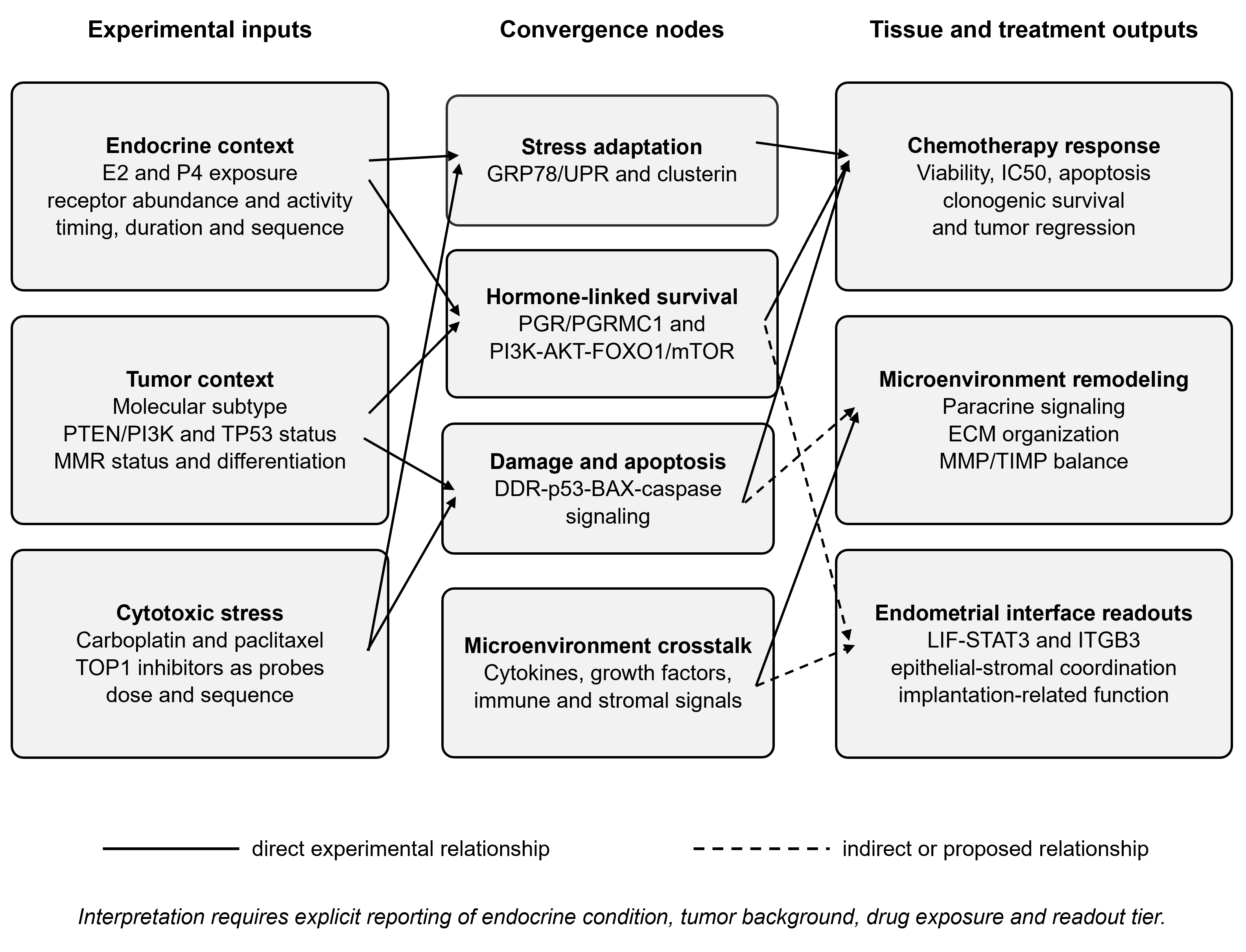
Fig. 1
Evidence-based framework for endocrine context and chemotherapy response in endometrial cancer. Endocrine exposure, tumor background, and cytotoxic stress converge on stress adaptation, hormone-associated survival, DNA-damage/apoptosis, and microenvironment pathways. Solid arrows indicate direct experimental evidence; dashed arrows indicate indirect or proposed relationships. LIF-STAT3 and ITGB3 are shown as endometrial-interface readouts rather than validated surrogates of chemotherapy response or fertility. DDR, DNA-damage response; E2, estradiol; ECM, extracellular matrix; P4, progesterone; PGR, progesterone receptor; UPR, unfolded protein response.
Three testable hypotheses follow. First, estrogen pretreatment will increase paclitaxel or platinum resistance only in tumors capable of inducing GRP78, clusterin, or functionally equivalent stress-buffering programs. Second, progesterone will have divergent effects according to classical progesterone-receptor isoform, PGRMC1 status, and AKT-FOXO1 activity. Third, endocrine conditioning may alter cytokine and MMP/TIMP outputs even when short-term drug sensitivity is unchanged. Testing these hypotheses requires factorial designs rather than separate hormone-only and drug-only experiments.
9. Limitations of the Review and of the Current Evidence
This review has methodological limitations that should be weighed when interpreting its conclusions. It is narrative rather than systematic: a single database was searched, screening and selection were performed by the authors without duplicate independent assessment, no validated risk-of-bias instrument was applied, and no quantitative synthesis was attempted. Restriction to full-text, peer-reviewed, English-language reports may have excluded relevant work, and the deliberate emphasis on mechanistically informative studies introduces a selection effect that cannot be quantified. The four evidence categories used here are an analytical device rather than a validated grading system, and other authors might reasonably classify individual studies differently.
The primary literature carries its own constraints. Most direct endocrine-chemotherapy evidence rests on a small number of two-dimensional cell lines, principally Ishikawa, ECC-1, KLE, and RL95-2, which do not represent the full molecular spectrum of endometrial carcinoma and which are commonly exposed to supraphysiologic hormone concentrations for short periods. Several key observations, including the MYH14-Wnt mechanism, currently depend on single reports without independent replication, and negative or null findings are likely to be underrepresented. Organoid and xenograft studies remain few, and no study identified here combined a defined endocrine condition, a clinically relevant carboplatin-paclitaxel schedule, and stromal or immune readouts within a single experimental system. Clinical corroboration is also absent: no trial has stratified chemotherapy outcome by hormone-receptor status together with the stress-adaptation or membrane-progesterone markers discussed above, and the routine addition of immune checkpoint inhibitors to chemotherapy introduces a further variable whose interaction with endocrine context has not been examined. The framework proposed here should therefore be read as a hypothesis-generating structure for study design rather than as a validated model.
10. Conclusion
The available evidence supports a qualified conclusion: endocrine-associated signaling can modify chemotherapy response in endometrial cancer, but the direction and magnitude of the effect depend on receptor context, molecular subtype, stress-adaptation pathways, drug class, and treatment sequence. Direct evidence is strongest for estrogen-GRP78, estrogen-clusterin, PGRMC1, AKT-FOXO1, and MYH14-Wnt mechanisms affecting paclitaxel, platinum, doxorubicin, or progesterone response. Evidence directly linking endocrine conditioning to camptothecin-family sensitivity is insufficient, so TOP1 inhibitors should be framed as mechanistic probes rather than the clinical center of the review.
Endometrial receptivity pathways and MMP/TIMP-mediated remodeling remain valuable physiologic comparators and potential tissue-interface readouts. They should not be treated as established proxies for tumor chemosensitivity or fertility outcomes. Future work should use molecularly characterized cell lines, patient-derived organoids, stromal co-cultures, and in vivo models with explicit endocrine timing and clinically relevant drug schedules. Such designs can determine when endocrine context changes intracellular drug response, when it primarily remodels the microenvironment, and when these effects extend to functional endometrial tissue outcomes.
Abbreviations
AKT, protein kinase B; BAX, BCL2-associated X protein; DDR, DNA-damage response; ECM, extracellular matrix; ER, estrogen receptor; FOXO1, forkhead box O1; GRP78, glucose-regulated protein 78; IC50, half-maximal inhibitory concentration; ITGB3, integrin beta 3; LIF, leukemia inhibitory factor; MAPK, mitogen-activated protein kinase; MMP, matrix metalloproteinase; MMR, mismatch repair; mTOR, mechanistic target of rapamycin; PARP, poly(ADP-ribose) polymerase; PGR, progesterone receptor; PGRMC1, progesterone receptor membrane component 1; PI3K, phosphatidylinositol 3-kinase; PRA and PRB, progesterone receptor isoforms A and B; PTEN, phosphatase and tensin homolog; SN-38, 7-ethyl-10-hydroxycamptothecin; STAT3, signal transducer and activator of transcription 3; TIMP, tissue inhibitor of metalloproteinases; TOP1, topoisomerase I; UPR, unfolded protein response.

